Specimen Imaging
Several specialized imaging technologies for both in vitro and non-biological specimens are available at the CMGI. High resolution X-ray microscopy allows analysis of internal structures of biological and non-biological specimens at a resolution of 1.5-20 μm. Specimen sizes up to 2 cm can be accommodated. A fluorescence imaging cryomicrotome provides high resolution (10-30 μm) fluorescence images of frozen tissue sections from rodent-sized specimens. A cryostat and phosphor imager are available for autoradiography, the gold standard for measuring biodistribution of radiolabeled molecules.
Further information is provided below.
MicroXCT-200 (Carl Zeiss X-ray Microscopy)
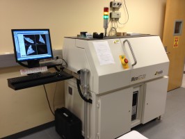 The MicroXCT-200 CT specimen scanner provides ultra high-resolution imaging in the 1.5 – 20 μm range. The high contrast 3D images allow visualization and analysis of fine structural features within samples as large as several cm. Both biological and non-biological samples may be imaged with this scanner. The MicroXCT-200 consists of a 90 kV micro-focused source; CCD camera; multiple optics to switch between resolution imaging modes; phase enhanced detectors; and a precision stage resting on a granite base. Three different detectors (based on a scintillator coupled to an optical light objective), with different magnification factors (4X, 10x and 20X), are installed on a software-controlled turret. X-rays are transmitted through the specimen and converted into optical photons by the scintillator. These optical photons are magnified by the optical light objective and captured by the CCD camera. The final resolution ranges from 1.5 µm to 20 µm depending on the magnification factor, with a sample field of view ranging from 1 – 50 mm.
Fluorescence Imaging Cryomicrotome (Barlow Scientific)
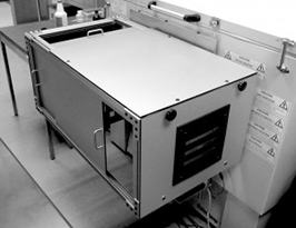 An imaging cryomicrotome is available for acquiring high resolution (10-30 μm) fluorescence images of frozen tissue sections up to rodent-sized specimens (maximum specimen size: 11 x 24 cm). This system consists of a light-tight cryomicrotome, a high intensity broad-band excitation source filtered by one of five computer-selectable filters on one wheel, a second wheel that filters the image with one of five computer-selectable filters, and CCD camera to record images. The excitation source emits over a broad spectrum up into the near infra-red and is thus suitable for in vivo studies with a range of fluorophores, quantum dots, and fluorescent proteins. The excitation light passes through an excitation filter wheel that illuminates the exposed face of a frozen block of tissue. The returning fluorescence from the specimen passes through the emission filter that selects the fluorescence wavelength of interest and a Nikon F-mount lens zoom lens. An Apogee U32 CCD camera with 2184×1472 pixels pixels (3 Mpixels) detector records each image. The system can image blocks as large as 5 x 5” (blockface surface) by 10” long. Image resolution is 18 microns/pixel at a 4 cm field of view. The microtome part of the system removes successive slices from the frozen specimen but instead of saving them, as in a conventional microtome, the slices are discarded. The system images the exposed face of the frozen specimen and proprietary software takes the images from successive slices to deduce the actual location of the fluorescence; the software takes into account that the fluorescence imaged originates from a range of depths into the specimen that depend on the tissue optical properties at the excitation and fluorescence wavelengths. Multiple images can be taken between slices. This procedure enables imaging of multiple fluorophores each on a separate image but all automatically registered.
Autoradiography
Autoradiography remains the gold standard for measuring in vivo biodistribution of radiolabeled molecules. This imaging technique requires tissue sectioning and imaging on film or phosphor screen. A cryostat (CM 1850, Leica) operated and maintained by CMGI staff is available for sectioning frozen tissue. Tissue samples are fixed and frozen in O.C.T. (Tissue-Tek, USA) by liquid nitrogen, cryogen spray, or a dry ice slurry. The microtome allows sectioning in thicknesses from 1-60 µm in increments of 1 µm (1-10 µm), 2 µm (10-20 µm) and 5 µm (20-60 µm). Maximum specimen size is 55 mm, which allows longitudinal sectioning of a whole mouse (up to approximately 25 g). Protocols for autoradiography, histological staining (H&E) and immunohistochemistry are available.
Phosphor Imager: Autoradiographic specimen imaging is performed with storage phosphor screens, rather than film, using a STORM 860 Phosphor Imager (GE Healthcare; formerly Amersham Biosciences). Two phosphor screens (35 x 43 cm) with an intrinsic resolution of 50 µm are available. ImageQuant software provides qualitative and quantitative data analysis.