X-Ray Computed Tomography (CT)
Benefits of MicroCT Imaging
• Good anatomical imaging of bone, tumor, vascular density (with appropriate contrast agent)
• Quantitative measurements
• High resolution relative to microPET
Ideally Suited For
• Anatomical imaging
• Lung imaging
• Multimodality imaging with microPET to combine functional and anatomical information in a single image
Not Suited For
• Functional imaging
Disadvantages
• Radiation dose to animal
• Poor soft tissue contrast without appropriate contrast agent
X-ray computed tomography (CT) provides high resolution 3-D anatomical imaging. It is particularly useful for imaging bone and fat, but can also provide capabilities for soft-tissue and vascular imaging by the use of suitable contrast agents. Animals can be scanned by PET and CT in the same session and the images co-registered to give a single image combining functional PET with anatomical CT data.
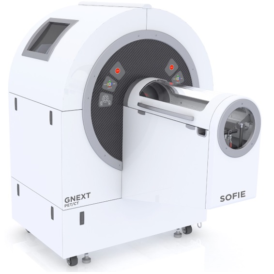 GNEXT PET/CT (Xodus Biosciences): Alignment and integration of the dedicated GNEXT CT component with the PET scanner allows for the acquisition of anatomical information that can be used for accurate image registration, attenuation correction, and anatomical localization of radiotracer uptake. The CT component can also operate as standalone, providing spatial resolutions down to 20 microns.