Magnetic Resonance Imaging (MRI)
Benefits of MRI
• Imaging provides both anatomical and functional information
• High spatial resolution
• Quantitative measurements
• No irradiation of animal
• Translational to clinic
Ideally Suited For
• Neuroscience and developmental biology studies
• Measuring blood oxygenation, water diffusion
• Assessing permeability, perfusion and vascular density
• Multimodality imaging with microPET to combine functional and anatomical information in a single image
Not Suited For
• Pharmacokinetic studies
Disadvantages
• Low sensitivity
• Fewer contrast agents than microPET, microSPECT
Magnetic resonance imaging (MRI) provides excellent soft-tissue contrast, allowing high-resolution anatomical imaging. More recently, the development of molecular-targeted contrast agents now allows high-resolution molecular imaging, which has increasingly been used for multimodal imaging in combination with PET to meld in a single image the exceptional anatomical resolution of MRI with the powerful functional imaging ability of PET. Research applications of MRI include stem cell and pathophysiologic imaging, including atherosclerosis, as well as volumetric brain imaging.
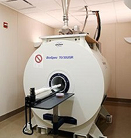 BioSpec 7T The CMGI is equipped with a Bruker BioSpec 7T horizontal bore system designed specifically for small animal imaging . Dedicated animal beds for mice (up to 35 g) and rats (up to 400 g) incorporate a circulating warm-water circuit to maintain body temperature, and can be fitted with a nose cone for anesthetic inhalation. A stereotactic frame is integrated into the animal beds to provide reproducible positioning of the same animal over multiple imaging sessions. Both cardiac and respiratory gating are available and may enhance image quality for some imaging protocols. Of special interest is the potential for PET/MRI multimodality imaging. A second generation PET insert being developed in Dr. Cherry’s laboratory will be fitted within the bore to obtain simultaneous data acquisition and a co-registered PET/MRI image. BioSpec 7T The CMGI is equipped with a Bruker BioSpec 7T horizontal bore system designed specifically for small animal imaging . Dedicated animal beds for mice (up to 35 g) and rats (up to 400 g) incorporate a circulating warm-water circuit to maintain body temperature, and can be fitted with a nose cone for anesthetic inhalation. A stereotactic frame is integrated into the animal beds to provide reproducible positioning of the same animal over multiple imaging sessions. Both cardiac and respiratory gating are available and may enhance image quality for some imaging protocols. Of special interest is the potential for PET/MRI multimodality imaging. A second generation PET insert being developed in Dr. Cherry’s laboratory will be fitted within the bore to obtain simultaneous data acquisition and a co-registered PET/MRI image. |
The following gradient/shim systems and coils are available:
- Gradient/shim system (BGA-20S OD: 301.5 mm; ID: 200.5 mm).
- Gradient/shim system (BGA-12S OD: 200 mm; ID: 116 mm).
- Circular polarized transmit/receive, or transmit only, coil (OD: 200 mm; ID: 154 mm) for rabbit body imaging. Used with the BGA-20S gradient.
- Linear transmitter/receive, or transmit only, coil (OD: 112 mm; ID: 72 mm) for mouse and rat body imaging. Used with the BGA-12S gradient.
- Circular polarized transmit/receive coil (OD: 89 mm; ID: 60 mm) for mouse and rat whole body imaging. Used with the BGA-12S gradient.
- Circular polarized transmit/receive coil (OD: 44 mm; ID: 23 mm) for mouse brain imaging.
- Rat brain phased array receive-only coil array.
- Mouse brain phased array receive-only coil array.
- Linearly polarized receive-only surface coils (10, 20 and 30 mm diameters).
- Linearly polarized transmit/receive 20 mm surface coil for 1H and 13C imaging
- Linearly polarized transmit/receive 20 mm surface coil for 1H and 31P
- Filters for 19F imaging. A 19F coil will be purchased in the near future.
The updated ParaVision 7.0 software package for acquisition and processing allows 2D or 3D imaging with processing features that include magnification, ROI analysis and geometrical measurement. Three CMGI staff members are trained to use the system and can provide assisted MRI scans upon request. In some cases, after sufficient technical and safety training, unassisted scanning by PIs or their staff will also be available.
Please inquire (cmgiimaging@ucdavis.edu) for further information.