CMGI Imaging Gallery
These images from CMGI studies highlight some of the many possible research applications of mPET, mCT, MRI, optical, and Cerenkov luminescence tomography (CLT) imaging, as well as specimen imaging (x-ray microscopy, optical cryomicrotome). Our facility has extensive experience with in vivo, ex-vivo and non-biological imaging.
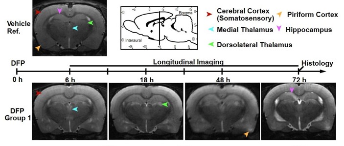
Brain injury at early time points after acute organophosphate exposure was assessed in a rat model. The organophosphate, diisopropylfluorophosphate [DFP]), was injected (4 mg/kg, s.c.) and in vivo MRI (Biospec 7T) was performed at multiple time points out to 72 hr post-DFP. T2-weighted (T2w), RARE transaxial images revealed prominent lesions in multiple brain regions, which varied in onset and progression. In the figure, lesions are identified by hyperintensity compared to adjacent tissue and to the vehicle reference (ref.) control scan. (Hobson BA, Rowland DJ, Supasai S, Harvey DJ, Lein PJ, Garbow JR. Neurotoxicology 66:170-78, 2018)
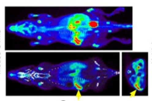
Renal cell carcinoma (RCC) has been variably sensitive and specific to image by PET using the traditional radiotracer, 18F-FDG, due to excretion into the urinary system. This study was designed to test whether 18F-Fluoroglutamine (18F-FGln) might be utilized for imaging RCC. In an orthotopic mouse model of RCC (left kidney), animals were imaged (Inveon DPET) for 30 min after a 15 min uptake of 18F-FGln injected i.v., followed immediately by Inveon CT imaging. Yellow arrow indicates tumor on left kidney, which was successfully imaged with 18F-FGln. Coronal and axial sections (bottom image) and maximum intensity projection image (top image) are overlaid on CT image. (Abu Aboud O, Habib SL, Trott J, Stewart B, Liang S, Chaudhari AJ, Sutcliffe J, Weiss RH. Cancer Res. 77:6746-58, 2017)
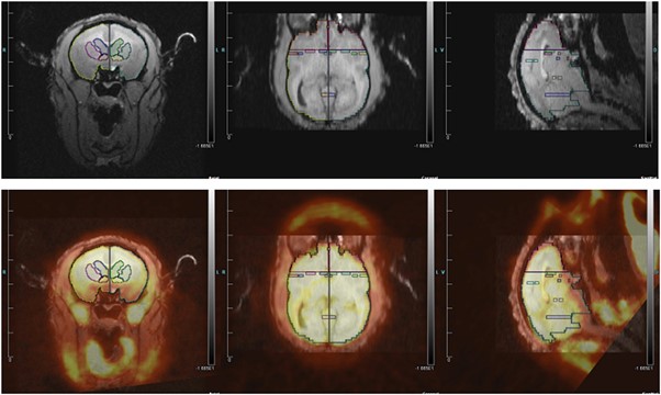
Titi monkeys form strong pair bonds characterized by selective preference for their pair mate and mate-guarding. During 30 min of 18F-FDG uptake (i.v.), caged males viewed either their female pair mate next to a stranger male (“jealousy” condition) or a stranger male with a stranger female (control condition). MicroPET images (mPET P4; 1 hr) (bottom row) were co-registered with MRI brain scans (top row, with regions of interest). 18F-FDG uptake was higher in several specific brain regions in the jealousy versus the control condition, similar to those involved in social exclusion in human studies. (Maninger N, Mendoza SP, Williams DR, Mason WA, Cherry SR, Rowland DJ, Schaefer T, Bales KL. Front Ecol Evol October 2017)
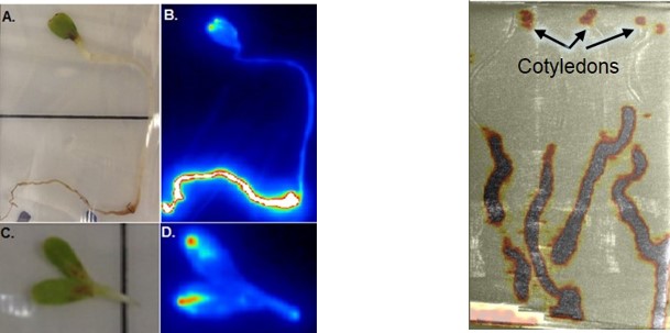
Engineered nanoparticles (NPs) are increasingly utilized in commercial products but their size makes it difficult to monitor their presence in the environment. This study assessed autoradiographic and PET/CT imaging of radiolabeled NPs as a means to detect the presence and track the movement of NPs in vivo in plants. The roots of lettuce seedlings were exposed to 64Cu-NPs, rinsed, and imaged by autoradiography (Storm 860 phosphoimager). Another group of 64Cu-NP – exposed seedlings were imaged by PET (15 min; Inveon DPET) followed by computed tomography (Inveon CT). Left: Autoradiograph after 2-hr root exposure to 64Cu-NPs; A. photo of seedling; B. autoradiograph of same seedling; C. photo of cotyledons; D. autoradiograph of same cotyledons. (color scale: white to dark blue, highest to lowest activity). Right: PET (DPET, 15 min)/CT image of 4 seedlings showing 64Cu-NPs in cotyledons and roots. (Davis RA, Rippner DA, Hausner SH, Parikh SJ, McElrone AJ, Sutcliffe JL. Environ Sci Technol. 51:12537-46, 2017)
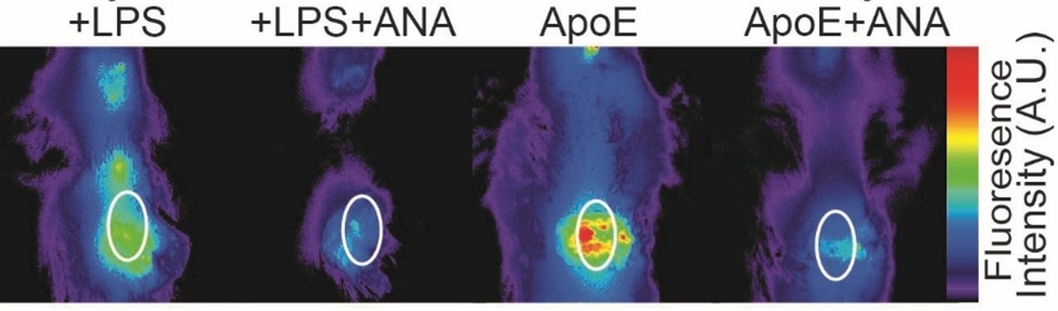
In mice with lipopolysaccharide (LPS) or atherosclerosis (ApoE)-induced inflammation, myocardial infarction was created and anakinra (ANA), an interleukin 1β inhibitor, was administered daily after 24 hours. Mice imaged in vivo (ProSense 750 FAST; Maestro 2) at 5 days post-MI showed reduced ProSense fluorescence in ANA-treated groups compared to untreated groups, indicating diminished inflammatory cell infiltration and activity. White ovals define heart location. (De Jesus NM, Wang L, Lai J, Rigor RR, Francis Stuart SD, Bers DM, Lindsey ML, Ripplinger CM. Heart Rhythm 14:727-36, 2017)

MR imaging in a rat model of acute organophosphate (OP) intoxication was used to assess persistence of neurological damage. Rats were imaged for 28 days post-OP intoxication. T2 weighted (top panel) and diffusion tensor images (DTI; bottom panel) were acquired on our BioSpec 7T and processed with Paravision. The colors in the DTI images represent direction of biased water diffusion in the brain. The orb in the upper right is a compass indicating water movement (fiber tract directionality): left-right is red, up-down is green, and in-out of the screen is blue. The corpus callosum (a tract of white matter connecting the left and right sides of the brain) is a bright, well defined yellow-red cord in the top third of the brain. Below that are swollen 1st and 2nd ventricles (symmetrical) 7 and 14 days after treatment, resulting from pooling of cerebrospinal fluid. (Hobson BA, Sisó S, Rowland DJ, Harvey DJ, Bruun DA, Garbow JR, Lein PJ. Toxicol Sci 157:342-53, 2017)
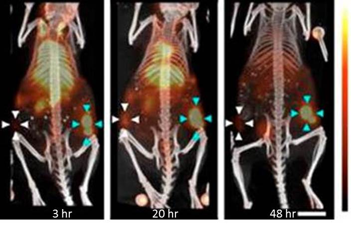
The potential for drug-carrying long-circulating liposomes (LCL) to enhance the therapeutic efficacy of magnetic resonance-guided focused ultrasound (MRgFUS) was investigated in a mouse breast cancer model. Following MRgFUS treatment (Bruker BioSpec 7T) of mammary carcinoma-bearing mice, 64Cu-LCL was administered (iv) and animals were imaged with PET-CT (mPET Focus 120 and Inveon CT) at 3, 20 and 48 hours. At each timepoint, US-treated tumors (blue arrows) had greatly increased uptake of 64Cu-LCL compared to contralateral controls (white arrows). The results suggest that MRgFUS in combination with doxorubicin-LCL may enhance therapy of unresectable tumors. (Wong AW, Fite BZ, Liu Y, Kheirolomoom A, Seo JW, Watson KD, Mahakian LM, Tam SM, Zhang H, Foiret J, Borowsky AD, Ferrara KW. J Clin Invest 126:99-111, 2016)
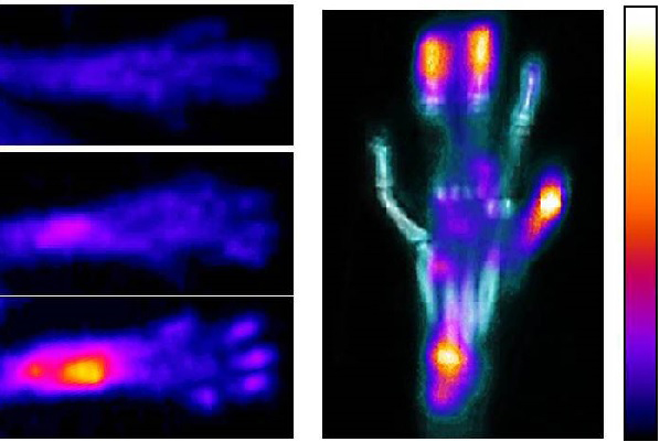
In a collagen-induced mouse model of inflammatory arthritis, which shares key pathological characteristics with human rheumatoid arthritis, the potential use of 18F-FDG PET/CT imaging to monitor and evaluate progression of the disease was explored. 18F-FDG PET and sequential CT scans were performed at day 0 (before first collagen injection; top image) and days 28 (middle image) and 56 (bottom and right). 18F-FDG uptake in paw joints correlated strongly with clinical and histological scores assigned independently at these time points. These results suggest that 18F-FDG PET/CT imaging in this mouse model may provide longitudinal, non-invasive assessment of the progression of autoimmune arthritic inflammation and of the impact of new therapeutic agents on the disease process. (Kundu-Raychaudhuri S, Mitra A, Datta-Mitra A, Chaudhari AJ, Raychaudhuri SP. Int J Rheum Dis 19:452-58, 2016)
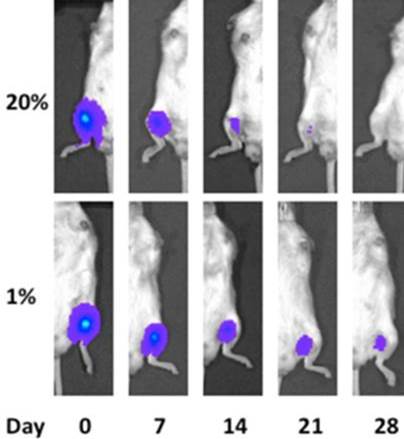
Survival of human mesenchymal stem cells/multipotent stromal cells (MSCs) following pre-exposure to hypoxia was tested in mice. Luciferase-expressing MSCs were pre-incubated for 48 hours in 1% oxygen (hypoxia) or 20% oxygen (normoxia) before injection into the medial hamstring muscle of NOD/SCID immune-compromised mice. Animals were imaged weekly (IVIS Spectrum; luciferin, ip, 10 min before 5 min exposure time). Cell survival and retention was significantly improved by hypoxia pretreatment at 14-28 days, suggesting that hypoxic pre-incubation of MSCs may improve therapeutic benefit. (Beegle JR, Magner NL, Kalomoiris S, Harding A, Zhou P, Nacey C, White JL, Pepper K, Gruenloh W, Annett G, Nolta JA, Fierro FA. Mol Ther Methods Clin Dev 33:1818-28, 2015).
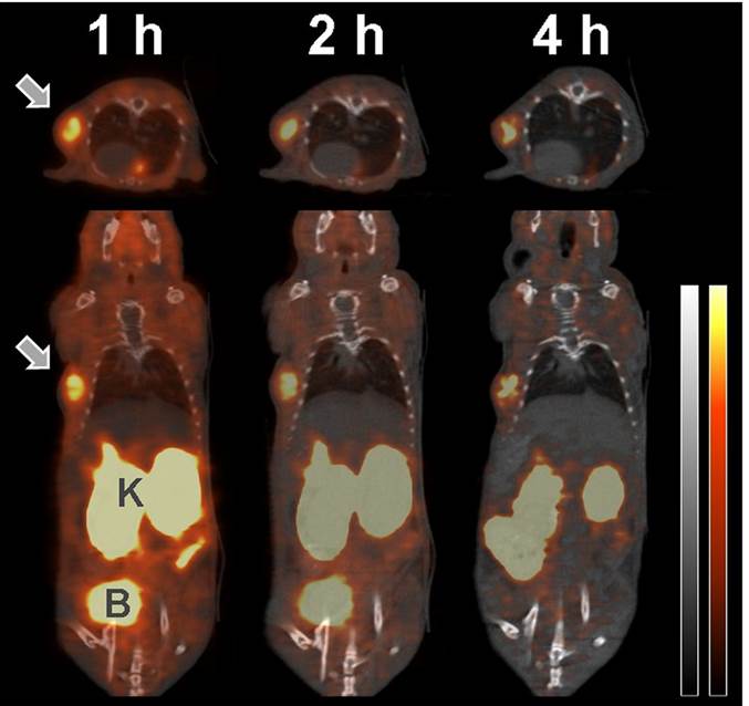
Pharmacokinetic profiles of several different PEGylated variations of the peptide, A20FMDV2, which selectively targets the cell surface receptor, integrin αvβ6, were assessed. The PET/CT image shows transaxial and coronal sections at 1, 2 and 4 hr after injection of 18F-FBA-PEG28-A20FMDV2-PEG28 (240 μCi, iv) in an athymic mouse model. The human pancreatic cancer cell tumor (BxPC-3 xenograf; shown by arrow) displayed high radiotracer uptake, resulting in favorable tumor-to-pancreas and tumor-to-muscle ratios. Autoradiography and immunochemistry confirmed colocalization of integrin αvβ6 expression and enhanced radioactivity. The authors concluded that bi-PEGylation may enhance uptake and retention of the peptide radiotracer in αvβ6-positive tumors. (PET – red; CT – grey; B – bladder; K- kidneys). (Hausner SH, Bauer N, Hu LY, Knight LM, Sutcliffe JL. J Nucl Med 56:784-90, 2015)
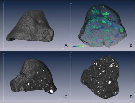
A fragment (0.5 g) of meteorite from the asteroid impact near Chelyabinsk, Russia in 2013 was analyzed for mineral content by high-resolution X-ray microscopy (MicroXCT-200). Imaging parameters: 85 kV, 93 mA, 8 sec exposure/projection, 2600 projections, 7.5 hr acquisition time; 5.5 mm pixel size. (A) Surface rendering showing fusion crust; (B) Volume rendering of internal composition: FeNi and FeS (green); FeNi and FeSi intermingled with silicates (light blue); (C) Single slice view; (D) Full section of the X-ray CT image. (Popova OP, Jenniskens P, Emel’yanenko V, et al. Science 342:1069-73, 2013)
") Image contrast, tumor volume and biodistribution were investigated to compare the standard clinical radiotracer, 18F-FDG, with long-circulating 64Cu-liposomes in a mouse mammary carcinoma model. Mice were imaged (Focus 120) 30 min after 18F-FDG (13.5 μCi/g, iv, 0.5 hr). Four hours later they received 64Cu-liposomes (8 and 16 μCi/g, iv) and were imaged at 6, 18, 24 and 48 hours. The 64Cu-liposomes accumulated in tumors (arrows) by 18 hours and remained elevated through 48 hours even as background activity diminished. 64Cu-liposomes produced tumor images with contrast and volume estimates comparable to tumors imaged with 18F-FDG. The authors concluded that radiolabeled nanoparticles may be clinically useful contrast agents to facilitate tumor detection, identification of tumor margins and therapeutic evaluation. (Wong AW, Ormsby E, Zhang H, Seo JW, Mahakian LM, Caskey CF, Ferrara KW. Am J Nucl Med Mol Imaging 3:32-43, 2013)
Image contrast, tumor volume and biodistribution were investigated to compare the standard clinical radiotracer, 18F-FDG, with long-circulating 64Cu-liposomes in a mouse mammary carcinoma model. Mice were imaged (Focus 120) 30 min after 18F-FDG (13.5 μCi/g, iv, 0.5 hr). Four hours later they received 64Cu-liposomes (8 and 16 μCi/g, iv) and were imaged at 6, 18, 24 and 48 hours. The 64Cu-liposomes accumulated in tumors (arrows) by 18 hours and remained elevated through 48 hours even as background activity diminished. 64Cu-liposomes produced tumor images with contrast and volume estimates comparable to tumors imaged with 18F-FDG. The authors concluded that radiolabeled nanoparticles may be clinically useful contrast agents to facilitate tumor detection, identification of tumor margins and therapeutic evaluation. (Wong AW, Ormsby E, Zhang H, Seo JW, Mahakian LM, Caskey CF, Ferrara KW. Am J Nucl Med Mol Imaging 3:32-43, 2013)
Ice cores collected from the bottom of ice-covered lakes in Antarctica were imaged by CT to analyze microbial colonies and inorganic composition (right side of photo is the upward direction). Top: photograph of the core. Bottom: Rendered volume with user-defined color and transparency mapped onto the CT scan (software developed through U.C. Davis KeckCAVES). White – sand grains and carbonate layers that precipitated within the microbial communities; Red – finer diffuse sediments and carbonate; Blue – microbial mat; Transparent – ice. (D. Sumner, T. Mackey; Earth and Planetary Sciences, U.C. Davis)
 This study investigated whether mice engineered to be null for the EDA segment of fibronectin, an alternatively spliced protein segment previously observed to be necessary for progression of inflammatory lung and heart injury, exhibit less acute inflammation after knee trauma. Mice with injured right knee and sham-injured left knee were imaged with combined PET/CT at 14 and 25 days. The image shows two mice imaged simultaneously, first by DPET (18F-FDG, i.v.) then CT. Left mouse: Fibronectin EDA mutant; Right: Wild-type; images overlaid using fiducial marker for registration. Uptake was routinely greater in injured versus uninjured knees and there was significantly less average 18FDG uptake in the injured knees of EDA-null compared to wild-type mice 14 days after knee injury. (J. Peters, Internal Medicine and D. Haudenschild, Orthopaedic Surgery, U.C.D.)
![]() The potential benefit of decellularized liver matrix (DLM) as a carrier for hepatocyte transplantation was assessed with bioluminescence imaging. DLM prepared from mouse livers and infused with immortalized human fetal hepatocytes (hFH) was implanted into the omentum of immunodeficient mice. Bioluminescence imaging to track engraftment of transplanted cells was achieved by transducing the hepatocytes with a lentiviral vector encoding the luciferase gene. At multiple time points, isoflurane-anesthetized mice were imaged (Xenogen IVIS 100) following ip luciferin injection. The figure shows bioluminescent images of the same mice over time with 3 modes of cellular transplantation: DLM implantation, splenic injection or omentum injection. The images reveal successful engraftment of hFH using DLM implantation compared to the other modes, as indicated by persistence of bioluminescent signal intensity for 8 weeks. (Zhou P, Lessa N, Estrada DC, Severson EB, Lingala S, Zern MA, Nolta JA, Wu J. Liver Transpl 17:418-27, 2011)
The potential benefit of decellularized liver matrix (DLM) as a carrier for hepatocyte transplantation was assessed with bioluminescence imaging. DLM prepared from mouse livers and infused with immortalized human fetal hepatocytes (hFH) was implanted into the omentum of immunodeficient mice. Bioluminescence imaging to track engraftment of transplanted cells was achieved by transducing the hepatocytes with a lentiviral vector encoding the luciferase gene. At multiple time points, isoflurane-anesthetized mice were imaged (Xenogen IVIS 100) following ip luciferin injection. The figure shows bioluminescent images of the same mice over time with 3 modes of cellular transplantation: DLM implantation, splenic injection or omentum injection. The images reveal successful engraftment of hFH using DLM implantation compared to the other modes, as indicated by persistence of bioluminescent signal intensity for 8 weeks. (Zhou P, Lessa N, Estrada DC, Severson EB, Lingala S, Zern MA, Nolta JA, Wu J. Liver Transpl 17:418-27, 2011)
 |
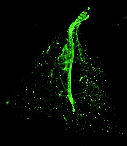 |
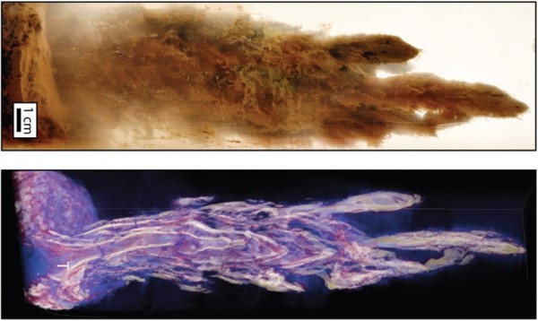
The distribution of inhaled particles was imaged with a fluorescence imaging cryomicrotome. Rats inhaled aerosolized fluorescent particles (2.5 mm mass median aerodynamic diameter) for 60 min and were necropsied. Frozen lung was sectioned by an imaging cryomicrotome (34 mm sections) and imaged in wavelengths to capture filled lumen and deposited particles separately. Left: white – airways; green – autofluorescence and fluorescence particles. Right: small green dots – inhaled particles; large structures – tissue autofluorescence. Deposited particles were identified in 2- and 3-dimensional images and were easily associated with lung structures. (A. Wexler, C. Wallis; Mechanical and Aerospace Engineering, U.C. Davis)
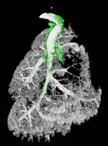
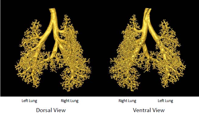 Rat lung casts imaged by microCT: Juvenile rats were exposed to ultrafine air pollutant particles to investigate potential effects of these pollutants on airway development. At 80-days of age, a cast of the lung’s conducting airways was prepared using a silicone injection technique. The lung cast was imaged with an InveonMM CT and a 3D image reconstructed for analysis of conducting airway bifurcations using custom software to quantify bifurcation generation number as well as length and diameter of conducting airways. These studies have shown that early-life exposure to ozone air pollutant results in significant persistent alterations in distal airway architecture that may have implications for the development of lung dysfunction in adult life.
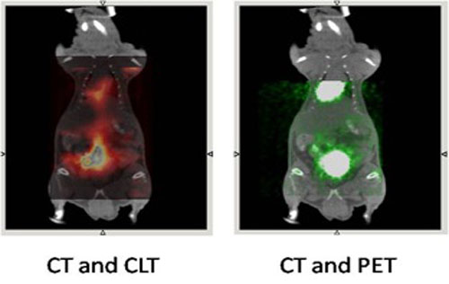 A study in 2009 reported the first use of Cerenkov radiation to optically image a mouse (Robertson R, Germanos MS, Li C, Mitchell GS, Cherry SR, Silva MD. Phys Med Biol 54:N355-65, 2009). Cerenkov radiation occurs when high energy particles such as positrons momentarily travel faster than light in tissues. The radiation is produced in a continuous spectrum from near ultraviolet through the visible spectrum and hence can be imaged using sensitive CCD methods optimized for optical imaging of small animals. In a further investigation, Cerenkov Luminescent Tomography (CLT) was used to noninvasively image the 18F-FDG concentration in a mouse. Briefly, the animal was injected with 18F-FDG (352 μCi) 2.5 hours before performing a 15-min PET scan (μPET II). MicroCT (Inveon SPECT/CT) and optical (Xenogen, IVIS 100) images were also acquired and the images overlaid. In the left figure, the colored regions are the optically imaged concentration of 18F-FDG reconstructed with CLT from the surface measurements of optical photons produced by Cerenkov radiation. The regions correspond to the heart and urinary bladder which characteristically display high 18F-FDG concentrations in the PET scan on the right (CT and PET). CLT is a new molecular imaging tool for preclinical studies that may provide a high-throughput, low cost PET alternative. Furthermore, it may be anticipated that the detection and utilization of Cerenkov radiation may lead to further advances in molecular imaging techniques.
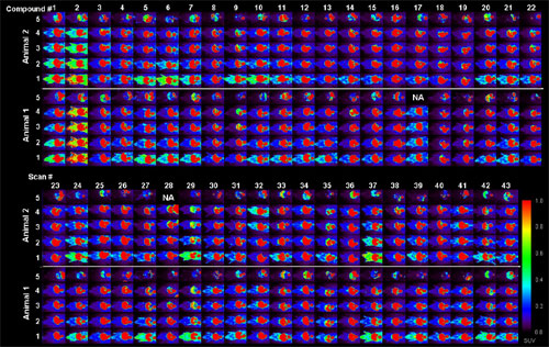 Multiple microPET (Focus 120) mouse images from a high-throughput screening study of [18F] labeled-peptides targeting the αvβ6 integrin. In vitro assays had identified 42 peptides with significant affinity and/or selectivity to αvβ6, and all compounds (+ 1 control) were evaluated in vivo with microPET on 11 consecutive days. Two mice were scanned simultaneously with each peptide (60 min dynamic scan post-injection and static scan at 180 min). A key finding was that the 4 most promising compounds identified in vivo did not correspond to the most promising compounds identified preliminarily in vitro. The image was awarded Siemens’ Most Innovative Preclinical Image of the Year, 2007. (Gagnon, MKJ, Marik J, Hausner SH, Abbey CK, Sutcliffe JL; High-throughput screening of molecular imaging agents using microPET. 54th Society of Nuclear Medicine Annual Meeting, Washington, D.C; June, 2007)
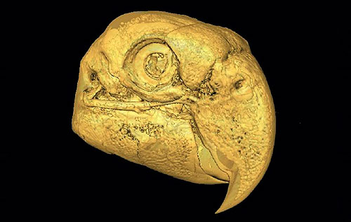
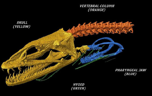 MicroCT (MicroCAT II) image of a moray eel partial skeleton of a preserved specimen. Bones of the skeleton were rendered by a surface rendering algorithm and false coloration was used to distinguish selected anatomical features. The investigator can use the surface mesh of the skull in a finite element analysis algorithm to study where forces are applied to the skull during biting in moray eels. This project uses 3D models generated from microCT scans to determine how variations in biting behavior of eels affects stresses on the skull, and how that may have affected the evolution of skull shape in both closely related as well as disparate lineages. (RS Mehta, Section of Evolution and Ecology, U.C.Davis)
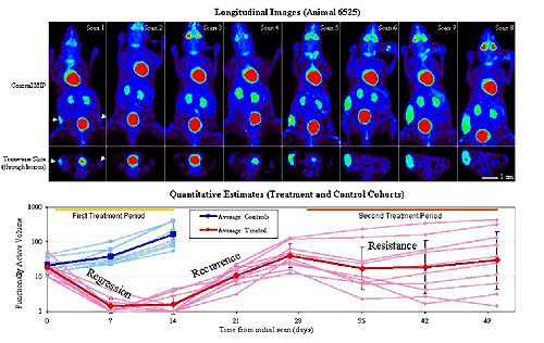 The inhibitory effect of the chemopreventative agent, rapamycin, on the development of a pre-malignant mammary lesion was assessed in a mouse model. Four weeks after receiving mammary tissue xenografts, animals were scanned (PET/FDG) for 8 successive weeks. Rapamycin was administered for 2 weeks, then stopped for 2 weeks, and finally readministered for 3 weeks. Coronal maximum intensity projections (MIP) of one animal, and a corresponding transverse slice through the mammary lesion (arrowhead), show lesion changes during the 8 weeks of imaging. The summary graph indicates that rapamycin was effective in reducing lesion size, but that cessation of treatment resulted in recurrence of lesions, which were more resistant to a second round of treatment with rapamycin. The study illustrates one of the advantages of non-invasive imaging by PET: use of a small number of animals in drug development studies to investigate both the primary question of therapeutic efficacy as well as secondary questions regarding 1) possible recurrence at completion of treatment and 2) efficacy during repeated treatments. (Abbey CK, Borowsky AD, Gregg JP, Cardiff RD, Cherry SR. J Mammary Gland Biol Neoplasia 11:137-49, 2006)
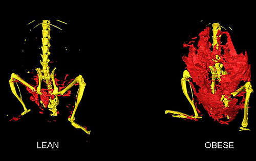 A pilot CT study (MicroCAT II) was performed to develop optimal image acquisition parameters and image processing techniques for measurement of adipose tissue in mice. Anesthetized mice were imaged with microCT (40 kVp, 400 uAmp, 800 ms projections, 201 projections) without a contrast agent. The segmented images depict adipose tissue (red) in relation to the skeleton (yellow) in lean and obese mouse models. Ex-vivo measurement of fat mass confirmed close agreement with the volume of adipose tissue imaged by microCT.
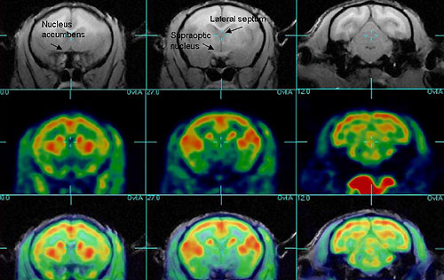 This project examined the neuroendocrine basis of social bonding in a primate model. Titi monkeys are monogamous and form strong emotional bonds between pair-mates. The PI used functional neuroimaging to investigate baseline differences in regional brain metabolism of pair-bonded and non-bonded animals. Conscious animals were administered 18F-FDG and were active for 35 min before being anesthetized for a 1-hr PET (microPET P4) brain scan. For MRI (1.5 Tesla), a 25-30 min brain scan was acquired. Image registration software (RView) was used to fuse each animal’s MRI and PET image to accurately identify anatomical regions and measure regional brain glucose metabolism. The figure shows selected image slices from MRI (top) and PET (center) scans in a pair-bonded male, and the coregistered images (bottom). Pair-bonded males showed lower relative glucose metabolism than lone males in all three of the areas identified on the MRI scan. (Bales KL, Mason WA, Catana C, Cherry SR, Mendoza SP. Brain Res 1184:245-53, 2007)
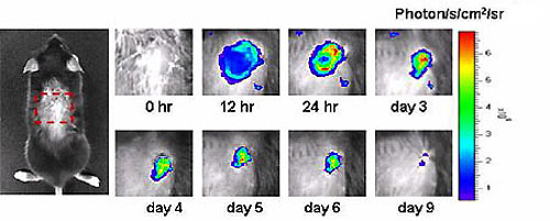 The kinetics of enhanced green fluorescence protein-polymorphonuclear leukocyte (EGFP-PMN) influx into a cutaneous wound site were investigated using fluorescence imaging. Transgenic mice, in which EGFP-PMN was knocked into the lysozyme gene, were imaged (Xenogen IVIS 100) at multiple time points after skin wounding. PMN influx was rapid during the first 12 hr and reached a maximum between 1 and 2 days, followed by a gradual decline over succeeding days. (Kim M-H, Liu W, Borjesson DL et al. J Invest Dermatol 128:1812-20, 2008)
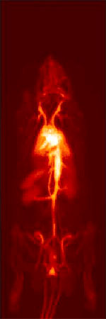 MicroPET (Focus 120) imaging was used to track the biodistribution of liposomes radiolabeled with 18F-fluorodipalmitin (18F-FDP) in a rat model. Liposome-encapsulated 18F-FDP, prepared on-site, was administered intravenously and imaged during a 90-min dynamic scan using continuous bed motion. Liposomes (average size = 45 nm) remained in the circulation at near-constant levels for the duration of the scan, providing a detailed map of the vasculature as seen in this maximum intensity projection image. Visualization of the lungs, liver, kidney and spleen reflect the vascular blood flow in these organs. Liposomes can be prepared with surface peptides or antibodies to target specific cell surface receptors. (Marik J, Tartis MS, Zhang H, Fung JY, Kheirolomoom A, Sutcliffe JL, Ferrara KW. Nucl Med Biol 34:165-71, 2007)
The formalin-preserved head of a macaw was imaged by microCT (MicroCAT II) as part of a pilot project to assess the feasibility of studying how the relationships between the individual bones of the skull and jaw are altered during opening and closing the beak. The image required 2 bed positions, and the acquired image was processed using an isosurface algorithm to provide a 3-dimensional impression of the bones of the skull and jaw.
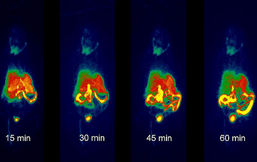 The biodistribution of the chemotherapeutic agent, paclitaxel, was studied by microPET (Focus 120) in a rat model. 18F-paclitaxel, prepared on-site, was administered intravenously and after 10 min the animal was dynamically imaged during a 60-min continuous bed motion scan. The time-segmented image set characterizes the biodistribution of free paclitaxel: once taken up by the liver it is solubilized in bile and secreted into the proximal small intestine, eventually entering the large intestine. (Ferrara lab)