Positron Emission Tomography (PET)
Benefits of MicroPET Imaging
- Isotopes of biologically important atoms can be substituted into targeting radiotracers allowing multiple applications
- Quantitative measurements
- Translational to clinic
Ideally Suited For
- Assessing cellular micro-environment (e.g., hypoxia) and metabolic changes, particularly in tumors, brain and heart
- Analyzing biodistribution of drugs and the dynamics of drug/receptor interactions
- Characterizing the stages and progression of a disease process and establishing signature biomarkers
- Assessing the efficacy of standard or experimental treatment modalities in small-animal models of human disease.
- Multimodality imaging with microCT or MRI to combine functional and anatomical information in a single image
Not Suited For
- Anatomical imaging
- High resolution imaging
Disadvantages
- Cyclotron needed for short-lived isotopes
- Moderate resolution (~1-1.5 mm)
- Radiation dose to animal
Positron emission tomography (PET) is a sensitive research tool that utilizes radiolabeled tracers for viewing molecular targets and biochemical pathways in vivo. The ability to image tissue perfusion, glucose metabolism, drug/receptor interactions, and gene expression have made it a particularly effective tool in oncology, cardiology and neuroscience research. The functional imaging achieved with PET can be combined with anatomical information obtained from the same animal using microCT or MRI to refine the identification of sites of PET radiotracer uptake.
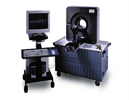
The nuclear imaging suite is equipped with 2 dedicated microPET scanners, an F120 (Siemens) and an Inveon DPET (Siemens), as well as an GNEXT PET/CT scanner (Xodus Biosciences). Three separate injection stations for each scanner are designed for simultaneous induction and/or maintenance anesthesia (isoflurane). Other support equipment include a dose calibrator (CRC 15R; Capintec), a commercial mouse tail illuminator/injection platform (Braintree Scientific), a warming light and both electric and circulating water heating pads. One PET scanner (PiPET, Brain Biosciences) is located at the California National Primate Research Center (CNPRC) and is operated and managed in partnership with the Multimodal Imaging Core of the CNPRC. This system is used exclusively for non-human primate studies (described below). The Multimodal Imaging Core at the CNPRC also operates a clinical PET/CT scanner (Discovery 610; GE) featuring high sensitivity, large field-of-view scanning.
 Inveon DPET This third generation small animal scanner uses LSO crystal elements for detection of gamma radiation produced from radioactive decay by positron emission. It features both high sensitivity (~ 10%) and high spatial resolution (1.4 mm FWHM with filtered back projection reconstruction). In addition, it is particularly well suited to whole body imaging of mice and rats: the bore is 12 cm, with an active transaxial field of view (FOV) of 10 cm and an axial FOV of 12.7 cm. Continuous bed motion can extend the axial FOV to 30 cm, providing a larger whole body single frame FOV for rats. Transmission scans with 57Co can be performed to apply attenuation correction during image reconstruction. The animal bed can be rapidly interchanged with the adjacent Inveon CT/SPECT bed to acquire CT scans of the same animal, thereby allowing reconstruction of a multimodality image that melds the functional information attained by PET within the anatomical framework rendered by CT.
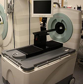
 Focus 120 (F120) The F120 is a third generation small animal PET scanner produced by Siemens Medical Solutions, Inc (formerly CTI Concorde Microsystems). Compared to the previous generation R4 model, which has the same ring diameter and axial length, the sensitivity and volumetric resolution are improved by more than 50% and 250%, respectively. The animal bed from the F120 can be easily removed and attached to our Inveon scanner to acquire CT anatomical images for co-registration with a PET image. Fiducial markers visible on both PET and CT are placed on or near the animal for precise co-registration of the images.
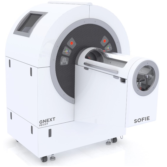 GNEXT PET/CT: The GNEXT PET/CT scanner is a state-of-the-art imaging system designed for small animal imaging. It offers several technical characteristics that contribute to its performance and utility in research applications. These include high spatial resolution (~1 mm isotropic), depth of interaction encoding that ensures uniform spatial resolution across the scanner field-of-view, 13% system sensitivity for photon detection, and greater than 10 cm axial field of view, enabling high geometric efficiency.

Discovery 610 PET/CT The Discovery 610 PET/CT (GE Healthcare) scanner, located at the CNPRC and operated and managed by the Multimodal Imaging Core, CNPRC, is a high sensitivity clinical scanner used primarily for whole-body imaging. Both hardware and software have been optimized to give enhanced sensitivity, allowing detailed imaging with lower radiation dose. The spatial resolution of the PET images is typically ~4-5 mm, and the CT images is ~ 1 mm. This combined PET/CT scanner is a powerful tool for assessing functional and anatomical information in a single fused image for research in non-human primates.
MicroPET Specifications Comparison
| MicroPET: | DPET | F120 | GNEXT | GE 610 (CNPRC) |
| Animal Port Diameter | 12 cm | 12 cm | 14 cm | 70 cm |
| Axial Field of View | 12.7 cm | 7.6 cm | 10 cm | NA |
| Absolute System Sensitivity | ~10% | 7% | 13% | ~10% |
| Reconstructed Spatial Resolution | ~ 1.5 mm | ~ 1.5 mm | ~1 mm | 4-5 mm (PET) ~ 1 mm (CT) |
| Reconstructed Volumetric Resolution | ~ 5 µL (FBP) | ~ 2 µL | NA |