Benefits of Fluorescence Imaging
• Multi-animal, high-throughput studies at relatively low cost
• Quantitative measurements
• High signal sensitivity
• Fluorescence probes, which can be activatable, are commercially available
• Multiple probes/targets can be imaged simultaneously
• No ionizing radiation
Ideally Suited For
• Cancer research, drug discovery and studies of inflammation, infectious diseases
Not Suited For
• High resolution imaging
Disadvantages
• Low resolution
• Tissue autofluorescence can increase background
• Less translational to clinic than microPET, microSPECT, MRI
• Depth of penetration of excitation and emitted light can be limiting
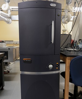 Ivis Spectrum: The IVIS Spectrum (Perkin Elmer) is a state-of-the-art high sensitivity optical imaging system for in vivo small animal studies using fluorescent reporter genes or injectable fluorescence contrast agents. All images are captured by a thermoelectrically cooled back-thinned, back-illuminated CCD camera, with a 2048 x 2048 array of 13.5 micron pixels (27 x 27 mm total area) and a 16-bit digitizer using 6-inch diameter optics with f-stop values that can be varied from f/1 to f/8. A computer-controlled filter wheel with 18 narrow-band (20 nm bandwidth) emission filters provide wavelength selectivity in the range 490-850 nm. A high intensity broad-band excitation source is filtered by one of ten computer-selectable excitation filters with bandwidths of 30 nm and covering an excitation range of 415-760 nm. The fluorescence light is delivered via an open fiber and can illuminate the subject in either reflective (epi-illumination) or transmission mode by selecting the appropriate fiber bundle switch. The system provides four levels of magnification ranging from high (20 micron) resolution (for single cell in vitro imaging) to a lower magnification, wide field setting, that allows imaging of 5 mice simultaneously.