Fluorescence Imaging Cryomicrotome (Barlow Scientific):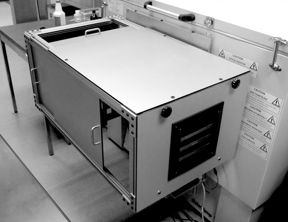
Fluorescence Imaging Cryomicrotome (Barlow Scientific):
An imaging cryomicrotome is available for acquiring high resolution (10-30 mm) fluorescence images of frozen tissue sections up to rodent-sized specimens (maximum specimen size: 11 x 24 cm). This system consists of a light-tight cryomicrotome, a high intensity broad-band excitation source filtered by one of five computer-selectable filters on one wheel, a second wheel that filters the image with one of five computer-selectable filters, and CCD camera to record images. The excitation source emits over a broad spectrum up into the near infra-red and is thus suitable for in vivo studies with a range of fluorophores, quantum dots, and fluorescent proteins. The excitation light passes through an excitation filter wheel that illuminates the exposed face of a frozen block of tissue. The returning fluorescence from the specimen passes through the emission filter that selects the fluorescence wavelength of interest and a Nikon F-mount lens zoom lens. An Apogee U32 CCD camera with2184×1472 pixels pixels (3 Mpixels) detector records each image. The system can image blocks as large as 5 x 5” (blockface surface) by 10” long. Image resolution is 18 microns/pixel at a 4cm field of view. The microtome part of the system removes successive slices ...
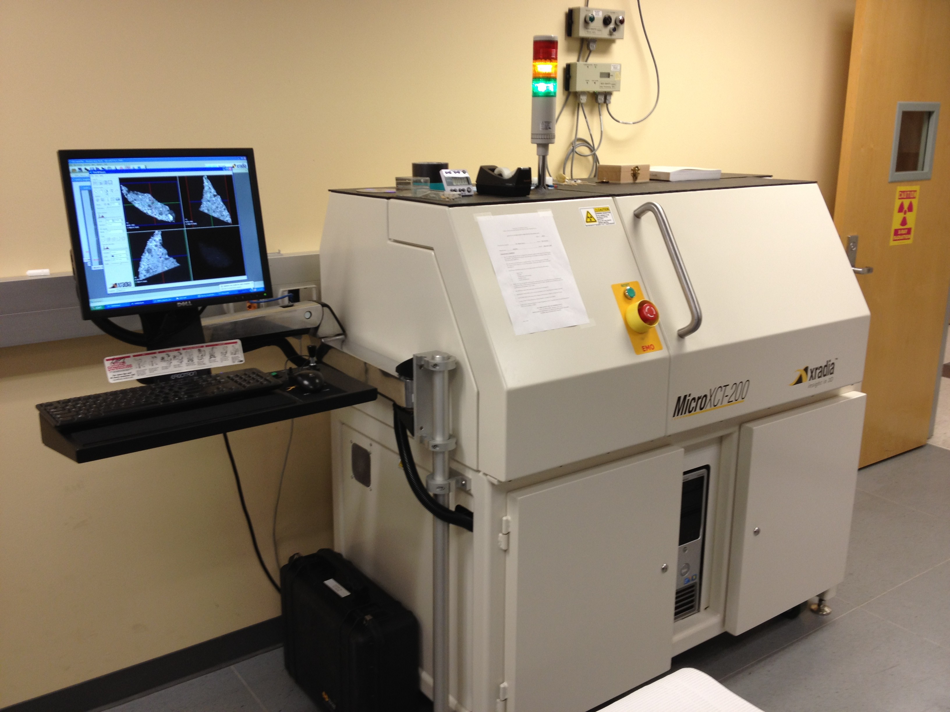 MicroXCT-200 (Carl Zeiss X-ray Microscopy): The MicroXCT-200 CT specimen scanner provides ultra high-resolution imaging in the 1.5 – 20 µm range. The high contrast 3D images allow visualization and analysis of fine structural features within samples as large as several cm. Both biological and non-biological samples may be imaged with this scanner. The MicroXCT-200 consists of a 90 kV micro-focused source; CCD camera; multiple optics to switch between resolution imaging modes; phase enhanced detectors; and a precision stage resting on a granite base. Three different detectors (based on a scintillator coupled to an optical light objective), with different magnification factors (4X, 10x and 20X), are installed on a software-controlled turret. X-rays are transmitted through the specimen and converted into optical photons by the scintillator. These optical photons are magnified by the optical light objective and captured by the CCD camera. The final resolution ranges from 1.5 µm to 20 µm depending on the magnification factor, with a sample field of view ranging from 1 – 50 mm.